Ultrasound Course Module 4
First and Second Trimester Scanning
Lesson 1: First Trimester Obstetrical Ultrasound
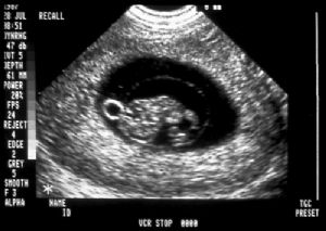 Yolk sac with fetus and incomplete fusion of amnion
and chorion at early gestation.
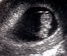
 Normal twin intrauterine pregnancy.
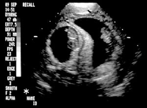
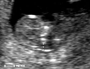 Normal nuchal translucency (NT) picture. Note calipers
on interior edge to interior edge of nuchal thickness.
- Guidelines (AIUM-ACR-ACOG-SMFM-SRU Practice Parameter for the Performance of Standard Diagnostic Obstetric Ultrasound Examinations, American Institute of Ultrasound, J Ultrasound Med 2018;9999:1-12.) For a complete list of guidelines see www.aium.org under above reference.
- Performance Objectives
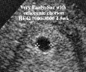
- Gestational sac/yolk sac measurements for presumptive pregnancy dating.
- May be visible by five menstrual weeks.
- Utilize three measurements: transverse, longitudinal and anteroposterior diameter to get a mean sac diameter (MSD). Example shown has a transverse diameter demonstrated at it shows the proper caliper position at inner edge of sac.
- May observe sac by 2-3 mm or 3-5 weeks. (de Crespigny LC, Cooper D, McKenna M. Early detection of intrauterine pregnancy with ultrasound. J Ultrasound Med 1988;7(1):7-10.)
- Ought to visualize an embryo at 25 mm gestational sac size.
- May be able to calculate gestational age in days (until sac diameter of 25 mm) by adding 30 to the MSD. So, MSD of 5 mm equals 5 weeks [or 35 days (30+5)]. (Nyberg DA, Filly RA, Filho DL, Laing FC, Mahoney BS. Abnormal pregnancy: Early diagnosis by US and serum chorionic gonadotropin levels. Radiology 1986;158(2):393-396.)
- The MSD less reliable after 14 mm (embryo of 5 mm).
- CRL with embryo located in uterus only certain way to rule out tubal pregnancy and obtain most accurate gestational age dating between 6-9 weeks. (Hadlock FP, Shah YP, Kanon DJ, Lindsey JV. Fetal crown-rump length: Reevaluation of relation to menstrual age (5-18 weeks) with high-resolution real-time US. Radiology 1992;182(2):501-5. & Pedersen JF. Fetal crown-rump length measurement buy ultrasound in normal pregnancy. Br J Obstet Gynaecol 1982;89(11):926-30.)
- Accuracy is 3-5 days for CRL. Note the proper measurement of the CRL from top of cranium to rump of fetus (not including the legs). (Robinson HP. Sonar measurement of fetal crown-rump length as a means of assessing maturity in the first trimester of pregnancy. BMJ 1973;4(5883):28-31. Robinson HP, Fleming JE. A critical evaluation of sonar "crown-rump length" measurements. Br J Obstet Gynaecol 1975;82(9):702-10. Drumm JE, Clinch J, MacKenzie G. The ultrasonic measurement of fetal crown-rump length as a method of assessing gestational age. Br J Obstet Gynaecol 1976;83(6):417-21.)
- Nuchal Thickness (NT)
- How Done
- Combine in nomogram with maternal serum analytes, maternal age, and gestational age to calculate risk
- Use ultrasound nuchal thickness at 10 6/7 weeks to 13 6/7 weeks (<3mm is normal)
- Maternal serum multiple analytes
- Pregnancy Associated Plasma Protein (PAPP-A)
- Free BhCG
- Results NT Testing
- Detects 91% of Downs
- Detects 97% of other Trisomies including Trisomy 18's
- Detects 40%+ of heart defects, skeletal syndromes
- Harbinger of IUFD, SGA, and miscarriage
- Number Fetuses
- Viability/FHR
- Adnexa/Uterine Anatomy
- Nicolaides KH, Azar G, Byrne D, Mansur C, Marks K. Fetal nuchal translucency: ultrasound screening for chromosomal defects in first trimester of pregnancy. BMJ 1992; 304: 867-869.
- Nicolaides KH, Brizot ML, Snijders RJ. Fetal nuchal translucency: ultrasound screening for fetal trisomy in the first trimester of pregnancy. Br J Obset Gynaecol 1994;101:782-786.
- Hyett JA, Perdu M, Sharland GK, Snijders RS, Nicolaides KH. Increased nuchal translucency at 10-14 weeks of gestation as a marker for major cardiac defects. Ultrasound Obstet Gynecol 1997;10:242-246.
- Snijders RJ, Noble P, Sebire N, Souka A, Nicolaides KH. UK multicentre project on assessment of risk of trisomy 21 by maternal age and fetal nuchal-tranlucency thickness at 10-14 weeks. Lancet 1998;352:343-346.
- Brady A, Pandya P, Yuksel B, Greeenough A, Patton M, Nicolaides KH. Outcome of chromosomally normal livebirth with increased fetal nuchal translucency at 10-14 weeks' gestation. J Med Genetics 1998;35:222-224.
- Hyett JA, Perdu M, Sharland GK, Snijders RS, Nicolaides KH. Using fetal nuchal translucency to screen for major congenital cardiac defects at 10-14 weeks of gestation: populaton based cohort study. BMJ 1999; 318:81-85.
- Zosmer N, Souter VL, Chan CSY, Huggon IC, Nicolaides KH. Early diagnosis of major cardiac defects in chromosally normal fetuses with increased nuchal translucency. Br J Obset Gynaecol 1999;106:829-833.
- Aitken DA, Wallace EM, Crossley JA, Swanston IA, Pareren Y, Maarle M, Groome NP, Macri JN, Conner MJ. Dimeric ihhibin A as a marker for Down's Syndrome in early pregnancy. N Engl J Med 1996;334: 1231-1234.