Ultrasound Course Module 5
Biometry, Prenatal Diagnosis, and Doppler
Lesson 3: Prenatal Diagnosis
- Cardinal Principle of Antenatal Detection
- Recognition of a departure from normal anatomy
- Absence of a normal structure
- Alteration of a normal structure (size, shape, location, continuity, echogenicity)
- Presence of an abnormal structure
- Reliability of Ultrasound Screening: Variables
- Prevalence of the condition
- Nature of the condition — "sonographic visibility"
- Quality of equipment and operator
- Imaging challenges:
- Maternal obesity
- Oligohydramnios
- Early/Late gestational age
- Fetal position
- Detection of Anomalies (Ewigman BG, Crane JP, Frigoletto FD, LeFevre ML, Bain RP, McNellis D. Effect of prenatal ultrasound screening on perinatal outcome. RADIUS Study Group. N Engl J Med. 1993 Sep 16;329(12):821-7. & Luck CA. Value of routine ultrasound scanning at 19 weeks: a four year study of 8849 deliveries. BMJ 1992;304(6840):1474-78.)
Sensitivity and SpecificitySensitivity % Sensitivity % n Radius 16.6 99.9 7685 Luck et. al. 84.3 98.6 8523 - Detection Rate by Classification of Anomalies (Romero R. Routine obstetric ultrasound. Ultrasound Obstet Gynecol 1993;3(5):303-07.)
- Lethal Anomalies 89%
- NICU Admission 77%
- Minor Anomalies 30%
- Most frequently missed:
- Cardiac malformations
- Cleft lip/palate
- Genitourinary (hypospadias, epispadius, ambiguous genitalia)
- Detection of Aneuploidy: "Soft Signs"
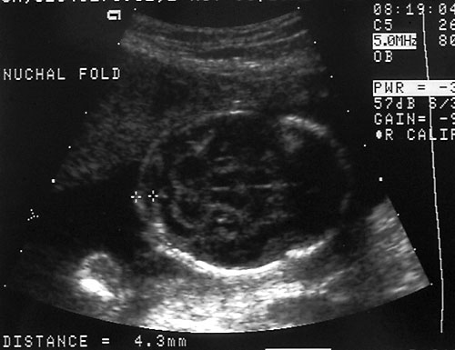
- Thickened nuchal skin fold
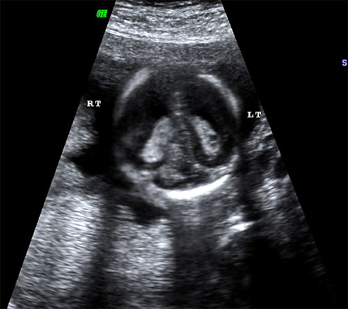
- Choroid plexus cysts
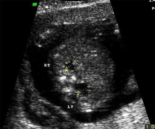
- Mild renal pyelectasis
- Hyperechoic bowel
- Echogenic intracardiac foci
- Short femur/humerus
- All are normal variants
- All are more prevalent in aneuploid fetus
Normal nuchal fold
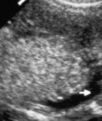
Choroid plexus cysts
Right sided choroids plexus cyst at arrow
Mild renal pyelectasis
Right pelvis is 4.2 mm and Left pelvis is 5.6 mm (normal < 4 mm)
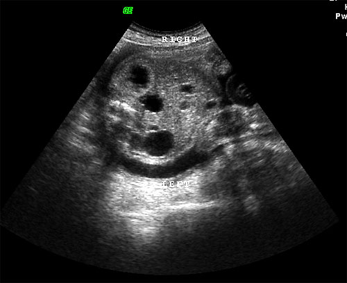
Mild renal pyelectasis
Severe right and left renal dilatation and dysplasia
Hyperechoic bowel
Hyperechoic bowel with same density as bone
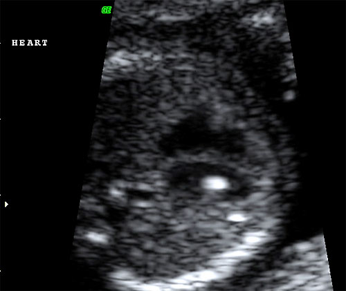
Echogenic intracardiac foci
Echogenic focus in left ventricle (bright spot in ventricle)
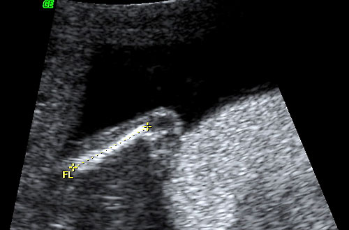
Short femur/humerus
Short femur as a "soft sign" for aneuploidy
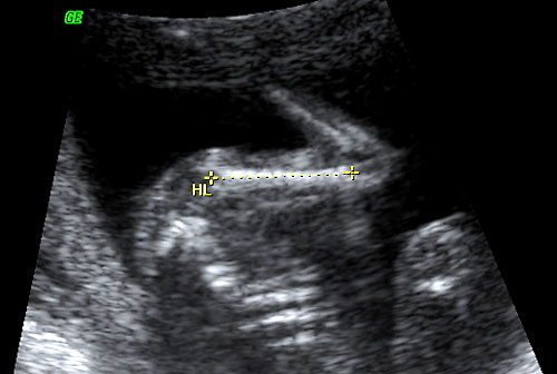
Short femur/humerus
Short humerus as a "soft sign" for aneuploidy - "Soft Signs" Scoring System (Bromley B, Shipp T, Benacerraf B. Genetic sonogram scoring index: accuracy and clinical utility. J Ultrasound Med 1999;8:523-28.)
- Two Points:
- Major structural anomaly
- Thickened nuchal fold
- One Point:
- Soft Signs o/t nuchal fold
- Two Points:
- "Soft Signs" Scoring System
Total Score 2 or greater:
Sensitivity (T21) — 85%
Sensitivity (T18 & T13) — 100%
PPV (T21):- Age > 40 — 7.1%
- Age 35-39 — 3.9%
- Age < 35 — 1.9%
- Nuchal Skin Fold (Benacerraf BR, Frigoletto FD Jr, Laboda LA. Sonographic diagnosis of Down syndrome in the second trimester. Am J Obstet Gynecol. 1985 Sep 1;153(1):49-52)
- Midtrimester (16-22 weeks)
- Edematous integument, with or without fluid collection
- Cut-off value: 6.0 mm